


See the metabolic markers behind your skin
One blood panel covers glucose, HbA1c, hsCRP, uric acid, and full lipids
- CLIA-certified labs
- HIPAA compliant
- Personalized health protocol
What are benign skin growths?
A benign skin growth is a noncancerous lump, bump, or discolored patch that forms when one type of skin cell multiplies in a contained way, staying put instead of invading nearby tissue or spreading elsewhere in the body. Clinicians may call the same thing a benign skin lesion or a non-malignant skin tumor, and the everyday version of the word is simply a growth.
Collecting a few of these over a lifetime is not a sign that something has gone wrong. It is closer to a default setting of aging skin: seborrheic keratoses alone are the most common benign epidermal tumor seen in dermatology practice. Two things are worth saying early and only once, because everything else rests on them. Most benign growths need no treatment at all, and only a clinician who has examined your skin can confirm that a particular growth is benign.
Benign growth vs. skin cancer: what the words mean
"Benign" describes behavior, not looks. A benign growth stays where it started, while a malignant one invades surrounding tissue and can travel to other parts of the body.
That distinction matters because appearance is a poor referee, and it fails in both directions. Seborrheic keratoses can mimic melanoma, squamous cell carcinoma, and basal cell carcinoma, and melanoma can imitate a seborrheic keratosis convincingly enough to fool an experienced eye. So it helps to know the features clinicians actually use, on the understanding that describing a pattern is not the same as clearing a lesion.
What causes benign skin growths?
Benign skin growths form when one population of skin cells is prompted to multiply locally: pigment cells, keratinocytes, blood vessels, fat cells, or fibrous tissue, each producing its own characteristic growth. The prompts fall into two broad groups, one internal and one external.
Age, genetics, and hormones
Age is the dominant driver, and the numbers are blunter than most people expect. Across an Australian population survey, seborrheic keratoses were found in 12% of 15 to 25 year olds and 100% of adults over 50, with the median count climbing from 6 lesions per person in the youngest group to 69 in those over 75. Their development is attributed to age and genetic predisposition. Hormones matter too: hyperinsulinemia drives keratinocyte and fibroblast proliferation through IGF-1 receptors, which is why certain growths cluster alongside metabolic change.
Sun exposure, friction, and skin injury
The external drivers are the ones you can partly see coming: light, rubbing, and repair. Seborrheic keratoses turn up more frequently per unit of surface area on sun-exposed sites. Friction leaves its own signature, which is why skin tags favor the neck, axilla, and groin, the places where skin meets skin or clothing all day. And repair itself can overshoot: keloid disorder is a benign fibroproliferative response to wound healing, with higher incidence in people with darker skin pigmentation.
The most common types of benign skin growths
The growths below are the ones adults most often find on themselves in a mirror. Each entry covers what the growth looks like, why it forms, and whether it usually needs anything done about it.
Common benign growth types at a glance
| Growth | Medical name | Typical appearance | Typical location | Usually needs treatment? |
|---|---|---|---|---|
| Mole | Melanocytic nevus | Usually under 5 mm, round or oval, evenly colored | Anywhere on the body | No |
| Seborrheic keratosis | Seborrheic keratosis | Waxy, "stuck-on" plaque | Anywhere except palms and soles, most often face and upper trunk | No, removal is cosmetic |
| Skin tag | Acrochordon | Small soft outgrowth on a narrow stalk | Neck, axilla, and groin | No, removal is elective |
| Cherry angioma | Cherry angioma | Small bright red papule | Front trunk most often, then the back | No |
| Lipoma | Lipoma | Soft, painless, mobile nodule under the skin | Most often the trunk | No |
| Sebaceous hyperplasia | Sebaceous hyperplasia | Yellowish papule with a dimpled center | Face | No, mainly cosmetic |
| Epidermoid cyst | Epidermoid cyst | 0.5 cm to several centimeters, central dark punctum | Face, neck, and trunk | Only if bothersome or ruptured |
| Milium | Milium | Tiny keratin-filled cyst | Face | No, usually self-limited |
| Dermatofibroma | Dermatofibroma | Firm nodule, typically 1 cm or less | Extremities | No |
| Keloid | Keloid | Raised overgrowth of scar tissue | Any healed wound or piercing site | Sometimes, to limit expansion |
Moles (melanocytic nevi)
A mole, or melanocytic nevus, is a cluster of pigment-producing cells. The baseline version is unremarkable by design: usually smaller than about 5 mm across, round or oval, with a smooth surface and a distinct edge, often dome-shaped, and an even pink, tan, or brown color. Only rarely does a common mole turn into melanoma.
Mole count is a different question from mole appearance. Having a lot of them is a risk marker rather than a diagnosis: an estimated 42% of melanomas are attributable to having 25 or more common moles, with risk rising about 1.7% per additional mole. Moles are not removed as a routine measure, and a mole that changes is a reason for a clinician to look rather than for you to decide.
Seborrheic keratoses
A seborrheic keratosis is a waxy, sharply bordered growth that looks stuck onto the skin rather than growing out of it, and it is the most common benign epidermal tumor in dermatologic practice. These appear anywhere except the palms and soles, most often on the face and upper trunk, and by age 50 essentially everyone has at least one. Removal is usually cosmetic, with no medical indication.
Skin tags (acrochordons)
A skin tag, or acrochordon, is a small soft outgrowth of skin on a narrow stalk, found in roughly 46% of the general population and rising with age. Tags collect on the neck, axilla, and groin, where they itch or catch on collars and straps.
The metabolic association is real but specific. Having more than five tags on the neck or axillae was associated with insulin resistance measured by HOMA-IR, and a HOMA-IR above 3.8 carried an odds ratio of 7.5. One tag on the eyelid says nothing of the sort. Removal is elective, driven by irritation or appearance.
Cherry angiomas
A cherry angioma is the most common vascular tumor of the skin, appearing as a small bright red papule that looks like a drop of paint. Both number and size increase with age, males carry more than females (median 16 versus 12), and the front trunk is the most common site, followed by the back.
The internet's favorite question about them deserves a precise answer. A link with cancer was suggested when these tumors were first described in the late 19th century but was never demonstrated. No treatment is required.
Lipomas
A lipoma is a benign tumor of fat cells sitting just under the skin, presenting as a soft, painless, mobile nodule, most often on the trunk and typically first noticed between the fourth and sixth decades of life. If you can slide it under your fingertips, that mobility is characteristic. Most require no treatment, and imaging is reserved for unusual sites or genuine diagnostic doubt.
Sebaceous hyperplasia
Sebaceous hyperplasia is an enlarged oil gland, showing up as a small yellowish papule with a dimple in the center, typically on the face and uncommon before adulthood. Prevalence reaches 10% to 16% in patients on long-term cyclosporine A. Treatment is optional, and no single technique is best: the choice depends on how many lesions there are, cost, skin phototype, age, and how much they bother the person living with them.
Epidermoid cysts and milia
An epidermoid cyst is a slow-growing, compressible, keratin-filled nodule beneath the skin, 0.5 cm to several centimeters across with a central dark opening called a punctum, most often on the face, neck, and trunk. A milium is a much smaller keratin-filled cyst, most often on the face, present in roughly half of full-term newborns and usually self-limited.
Squeezing an epidermoid cyst works against you on both counts. A ruptured cyst closely resembles a boil, with tenderness, redness, and swelling, and definitive treatment is complete excision with the cyst wall intact, which is harder once the wall has been broken.
Dermatofibromas and keloids
A dermatofibroma is a firm nodule usually 1 cm or less, most often on the extremities, most frequent between ages 20 and 50 and especially in women, with multiple lesions in about 10% of cases. Press the skin either side of one and the center dimples inward, a quirk of how the lesion tethers to the tissue below.
A keloid is a benign fibroproliferative overgrowth of scar tissue, with higher incidence in groups with darker skin pigmentation and associations with HLA, NEDD4, and VDR variants. The honest limit is that no current genetic test can predict keloid risk, and treatment aims at limiting scar expansion rather than resolving a fully understood mechanism.
Birthmarks and vascular growths
Birthmarks are growths present at birth or appearing in the first weeks of life, most commonly pigmented or vascular in origin, and they follow a different course and a different care pathway than the growths adults accumulate later.
Infantile hemangioma is the clinically important example. Most can be managed conservatively without intervention, but those that do need treatment should have specialist review ideally before 5 weeks of age. Propranolol is described as the standard systemic therapy, with topical timolol or pulsed dye laser used for small superficial lesions.
That early window is the whole point: a parent watching an infant birthmark grow or ulcerate has a reason to raise it with a clinician quickly rather than settle into watch-and-wait.
How to tell a benign growth from something suspicious
The features below describe patterns clinicians use, and none of them confirms that a growth is benign. They tell you whether a growth deserves a professional look.
Benign-looking features vs. concerning features
| Feature | Benign-leaning | Concerning |
|---|---|---|
| Symmetry | Round or oval, symmetrical | One half unlike the other |
| Border | Smooth surface with a distinct edge | Irregular, scalloped, or poorly defined |
| Color | Even pink, tan, or brown | Color varies within the spot |
| Size | Usually smaller than about 5 mm | Usually greater than 6 mm at diagnosis, though sometimes smaller |
| Change over time | Stable in size, shape, and color over months | Changing in size, shape, or color |
| Bleeding or crusting | Absent without injury | Bleeds or crusts without injury, or will not heal |
| How it compares | Resembles your other growths | Looks different from the rest |
The features that point to benign
Read down the left column and a pattern emerges: growths that are symmetrical, evenly colored, stable in size and appearance across months, and not bleeding or crusting are the ones clinicians associate with benign lesions. That is the profile of an ordinary common mole, and most of what you find on yourself will fit it.
The trouble is the exceptions, and they are not rare enough to ignore. Seborrheic keratosis-like melanoma and true seborrheic keratosis are often indistinguishable even to experienced dermatologists, because the lesion carries melanoma features and seborrheic keratosis features at the same time. Sebaceous hyperplasia counts basal cell carcinoma among its primary differential diagnoses. And fibroepithelioma of Pinkus, a basal cell carcinoma subtype, mimics a skin tag. A benign-looking pattern narrows the odds; it does not clear the lesion.
The ABCDE warning signs and other red flags
For pigmented lesions, there is a specific checklist worth knowing by heart. The five criteria are defined by the letters A through E:
- Asymmetry. One half of the spot does not match the other.
- Border. The edge is irregular, scalloped, or poorly defined.
- Color. The color varies from one area to another within the spot.
- Diameter. Melanomas are usually larger than 6 mm at diagnosis, though they can be smaller.
- Evolving. The spot looks different from the rest, or is changing in size, shape, or color.
For growths that carry no pigment, the red flags are similar in spirit: a growth that bleeds or crusts without injury, one that will not heal, and one that changes in size, shape, or color.
Now the part the checklist posters leave off. The more ABCDE signs a lesion showed, the more likely a non-dermatologist was to suspect it, rising from 8% with no signs to 32% with all five, yet the signs were not associated with catching melanoma at the in situ stage, and relying on ABCDE alone may miss early melanoma. Treat it as a prompt to get checked, never as a way to clear yourself.
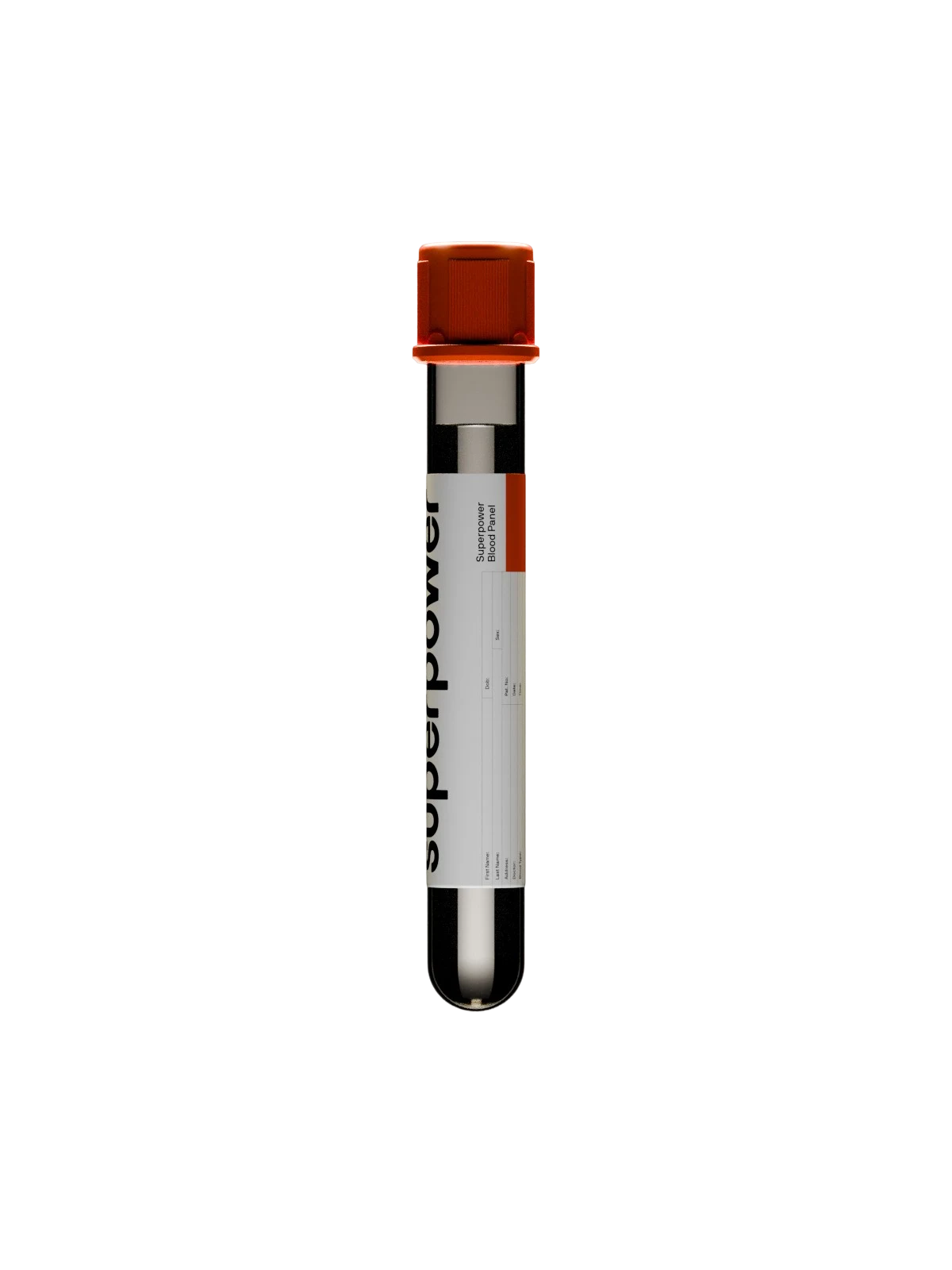
Test 100+ biomarkers
One blood draw. A full picture of your health, explained in plain language.
Test 100+ biomarkers
One blood draw. A full picture of your health, explained in plain language.
How dermatologists diagnose a skin growth
Diagnosis starts with a clinical examination of the growth and the skin around it, and for most benign growths that examination settles the question, with dermatoscopic or histologic assessment added when it does not.
Dermoscopy is where the gap between a trained look and an untrained one gets wide. A handheld magnifier with polarized light reveals structures the naked eye cannot resolve, and its use increases sensitivity for skin cancer detection, reduces the number of benign lesions biopsied per malignancy found, and allows thinner melanomas to be diagnosed. Those structures are surprisingly discriminating: shiny white structures and pseudopods carried odds ratios of about 6.7 for melanoma, with pseudopods 97.3% specific, although individual features had modest sensitivity on their own.
When the examination still leaves doubt, a biopsy takes the decision out of the realm of appearance and lets the tissue answer. Photo-based review has its place as triage rather than as a substitute: face-to-face assessment remained more accurate than remote review, at 67% to 85% agreement with the reference standard versus 51% to 85%, while remote review consistently cut waiting times.
Treatment and removal options for benign skin growths
Most benign skin growths need no treatment, and removal is usually chosen for comfort or appearance rather than medical necessity. When someone does want a growth gone, the techniques fall into a short and well-established list:
- Cryotherapy. Freezing the lesion with liquid nitrogen.
- Electrodesiccation. Destroying tissue with an electric current.
- CO2 laser. Vaporizing superficial lesions with focused light.
- Er:YAG laser. A shallower ablative laser used for surface growths.
- Shave excision. Removing a raised lesion flush with the skin.
- Snip excision. Cutting stalked lesions such as skin tags at the base.
- Surgical excision. Removing the whole lesion, and for epidermoid cysts taking the cyst wall out intact, with the procedure delayed if the cyst is actively infected.
Head-to-head data is thinner than you would hope, but it exists. In a randomized within-patient trial of 30 patients, each treated on four similar facial seborrheic keratoses with a different method, healing was significantly better with CO2 laser, Er:YAG laser, and electrodesiccation than with cryotherapy, and patient satisfaction was significantly lower with cryotherapy, while post-treatment pigmentation and texture did not differ significantly between the groups.
The selection principle is less about the machine than the person: effectiveness depends on lesion number, cost, skin phototype, age, and how much the growths bother you. One caution worth stating plainly. Removing a growth belongs with a clinician who has examined it, because at-home removal destroys the exact tissue that would otherwise have supplied the diagnosis.
When to see a doctor about a skin growth
See a clinician about any skin growth that is new, changing, bleeding, painful, or simply different from the others on your body, and about any growth you are unsure of, because appearance alone does not settle the question.
The specific escalation triggers are worth memorizing: a change in size, shape, or color; bleeding or crusting without injury; a lesion that will not heal; and any pigmented lesion meeting ABCDE criteria. Each of those is a prompt to get checked rather than a self-assessment you can complete at the bathroom mirror.
Some people warrant a lower threshold. Melanoma risk climbs with mole count, and an estimated 42% of melanomas are attributable to having 25 or more common moles, while the attributable fraction at low mole counts is very small. Infants with a growing or ulcerating birthmark belong in the same lower-threshold group, given the pre-5-week specialist review window. This article describes patterns; it is not a substitute for a clinician examining your skin, and nothing in it can determine whether a specific growth is benign.
How to monitor your skin at home
Self-checks are worth doing and easy to overtrust. The sensitivity of skin self-examination is low and highly variable, reported between 25% and 93%, while specificity runs higher at 83% to 97%, and education improves recognition of early melanoma signs. Put bluntly: a growth that looks stable to you has not been cleared.
A workable routine looks like this:
- Check your whole skin surface in good light, including the back, scalp, and the places you cannot see easily, using a mirror or another person's eyes.
- Photograph any growth you are tracking, with a ruler or a coin in frame for scale.
- Compare against that same photograph roughly every three months instead of relying on memory.
- Note anything new, changing, bleeding, or asymmetric.
- Book a clinician review for anything on that list rather than waiting to see whether it changes further.
Check the metabolic markers behind skin growths with a Superpower blood panel
Some benign skin growths sit alongside changes you can measure in blood. People with skin tags showed significantly higher HOMA-IR, fasting glucose and insulin, HbA1c, hsCRP, uric acid, total cholesterol, LDL, and triglycerides than controls, with 53.9% overweight and 33.6% obese. A skin finding like that is a reason to look at the metabolic picture underneath it, not a diagnosis in itself, and the growth itself still belongs with a dermatologist.
That is the part Superpower can help with. The Superpower Blood Panel covers many of those metabolic and inflammatory markers, including HbA1c, glucose, hsCRP, uric acid, and a full lipid panel, from a single annual draw. Superpower testing does not detect, screen for, rule out, or diagnose skin cancer or any skin growth.
Frequently Asked Questions
References
- Barthelmann S, Butsch F, Lang BM, Stege H, Großmann B, Schepler H, Grabbe S (2023). Seborrheic keratosis. *Journal der Deutschen Dermatologischen Gesellschaft*, *21*(3), 265–277. https://doi.org/10.1111/ddg.14984
- Moscarella E, Brancaccio G, Briatico G, Ronchi A, Piana S, Argenziano G (2021). Differential diagnosis and management on seborrheic keratosis in elderly patients. *Clinical, Cosmetic and Investigational Dermatology*, *14*, 395–406. https://doi.org/10.2147/CCID.S267246
- Yeatman JM, Kilkenny M, Marks R (1997). The prevalence of seborrhoeic keratoses in an Australian population: does exposure to sunlight play a part in their frequency? *British Journal of Dermatology*, *137*(3), 411–414.
- González-Saldivar G, Rodríguez-Gutiérrez R, Ocampo-Candiani J, González-González JG, Gómez-Flores M (2017). Skin manifestations of insulin resistance: from a biochemical stance to a clinical diagnosis and management. *Dermatology and Therapy*, *7*(1), 37–51. https://doi.org/10.1007/s13555-016-0160-3
- Belgam Syed SY, Lipoff JB, & Chatterjee K (2023). *Acrochordon*. StatPearls Publishing.
- James AJ, Torres-Guzman RA, Chaker SC, Sigel ME, Perdikis G, Supp DM, Dale Slater EL (2024). Global insights into keloid formation: an international systematic review of regional genetic risk factors and commonalities. *Wound Repair and Regeneration*, *32*(5), 746–757. https://doi.org/10.1111/wrr.13203
- National Cancer Institute (2022). *Common moles, dysplastic nevi, and risk of melanoma*. National Institutes of Health.
- Betz-Stablein B, Koh U, Edwards HA, McInerney-Leo A, Janda M, Soyer HP (2022). Anatomic distribution of cherry angiomas in the general population. *Dermatology*, *238*(1), 18–26. https://doi.org/10.1159/000517172
- Kolb L, Yarrarapu SNS, Robson O, & Rosario-Collazo JA (2026). *Lipoma*. StatPearls Publishing.
- Farci F, & Rapini RP (2023). *Sebaceous hyperplasia*. StatPearls Publishing.
- Zito PM, & Scharf R (2023). *Epidermoid cyst*. StatPearls Publishing.
- Gallardo Avila PP, & Mendez MD (2023). *Milia*. StatPearls Publishing.
- Myers DJ, & Fillman EP (2024). *Dermatofibroma*. StatPearls Publishing.
- Olsen CM, Carroll HJ, Whiteman DC (2010). Estimating the attributable fraction for cancer: a meta-analysis of nevi and melanoma. *Cancer Prevention Research*, *3*(2), 233–245. https://doi.org/10.1158/1940-6207.CAPR-09-0108
- Tamega AA, Aranha AM, Guiotoku MM, Miot LDB, Miot HA (2010). Association between skin tags and insulin resistance. *Anais Brasileiros de Dermatologia*, *85*(1), 25–31. https://doi.org/10.1590/S0365-05962010000100003
- Pastor-Tomás N, Bañuls J, Nagore E (2023). Clinical relevance of cherry angiomas. *Actas Dermo-Sifiliográficas*, *114*(3), T240–T246. https://doi.org/10.1016/j.ad.2022.10.040
- Hussein L, Perrett CM (2021). Treatment of sebaceous gland hyperplasia: a review of the literature. *Journal of Dermatological Treatment*, *32*(8), 866–877. https://doi.org/10.1080/09546634.2020.1720582
- Sebaratnam DF, Rodríguez Bandera AL, Wong LF, Wargon O (2021). Infantile hemangioma. Part 2: management. *Journal of the American Academy of Dermatology*, *85*(6), 1395–1404. https://doi.org/10.1016/j.jaad.2021.08.020
- American Academy of Dermatology (n.d.). *What to look for: ABCDEs of melanoma*. American Academy of Dermatology Association.
- Janowska A, Oranges T, Iannone M, Romanelli M, Dini V (2021). Seborrheic keratosis-like melanoma: a diagnostic challenge. *Melanoma Research*, *31*(5), 407–412. https://doi.org/10.1097/CMR.0000000000000756
- Duarte AF, Sousa-Pinto B, Azevedo LF, Barros AM, Puig S, Malvehy J, Haneke E, Correia O (2021). Clinical ABCDE rule for early melanoma detection. *European Journal of Dermatology*, *31*(6), 771–778. https://doi.org/10.1684/ejd.2021.4171
- Wolner ZJ, Yélamos O, Liopyris K, Rogers T, Marchetti MA, Marghoob AA (2017). Enhancing skin cancer diagnosis with dermoscopy. *Dermatologic Clinics*, *35*(4), 417–437. https://doi.org/10.1016/j.det.2017.06.003
- Williams NM, Rojas KD, Reynolds JM, Kwon D, Shum-Tien J, Jaimes N (2021). Assessment of diagnostic accuracy of dermoscopic structures and patterns used in melanoma detection: a systematic review and meta-analysis. *JAMA Dermatology*, *157*(9), 1078–1088. https://doi.org/10.1001/jamadermatol.2021.2845
- Finnane A, Dallest K, Janda M, Soyer HP (2017). Teledermatology for the diagnosis and management of skin cancer: a systematic review. *JAMA Dermatology*, *153*(3), 319–327. https://doi.org/10.1001/jamadermatol.2016.4361
- Zaresharifi S, Robati RM, Dadkhahfar S, Forouzanfar MM, Zaresharifi N (2021). Efficacy and safety of cryotherapy, electrodesiccation, CO2 laser, and Er:YAG laser in the treatment of seborrheic keratosis. *Dermatologic Therapy*, *34*(5), e15083. https://doi.org/10.1111/dth.15083
- Hamidi R, Peng D, Cockburn M (2010). Efficacy of skin self-examination for the early detection of melanoma. *International Journal of Dermatology*, *49*(2), 126–134. https://doi.org/10.1111/j.1365-4632.2009.04268.x
- Sari R, Akman A, Alpsoy E, Balci MK (2010). The metabolic profile in patients with skin tags. *Clinical and Experimental Medicine*, *10*(3), 193–197. https://doi.org/10.1007/s10238-009-0086-5
























 Heart & Vascular Health
 Metabolic Health
 Energy
 Liver Health
 Kidney
 Sex Hormones
 Thyroid Health
 Nutrients
 Immune System
 Inflammation