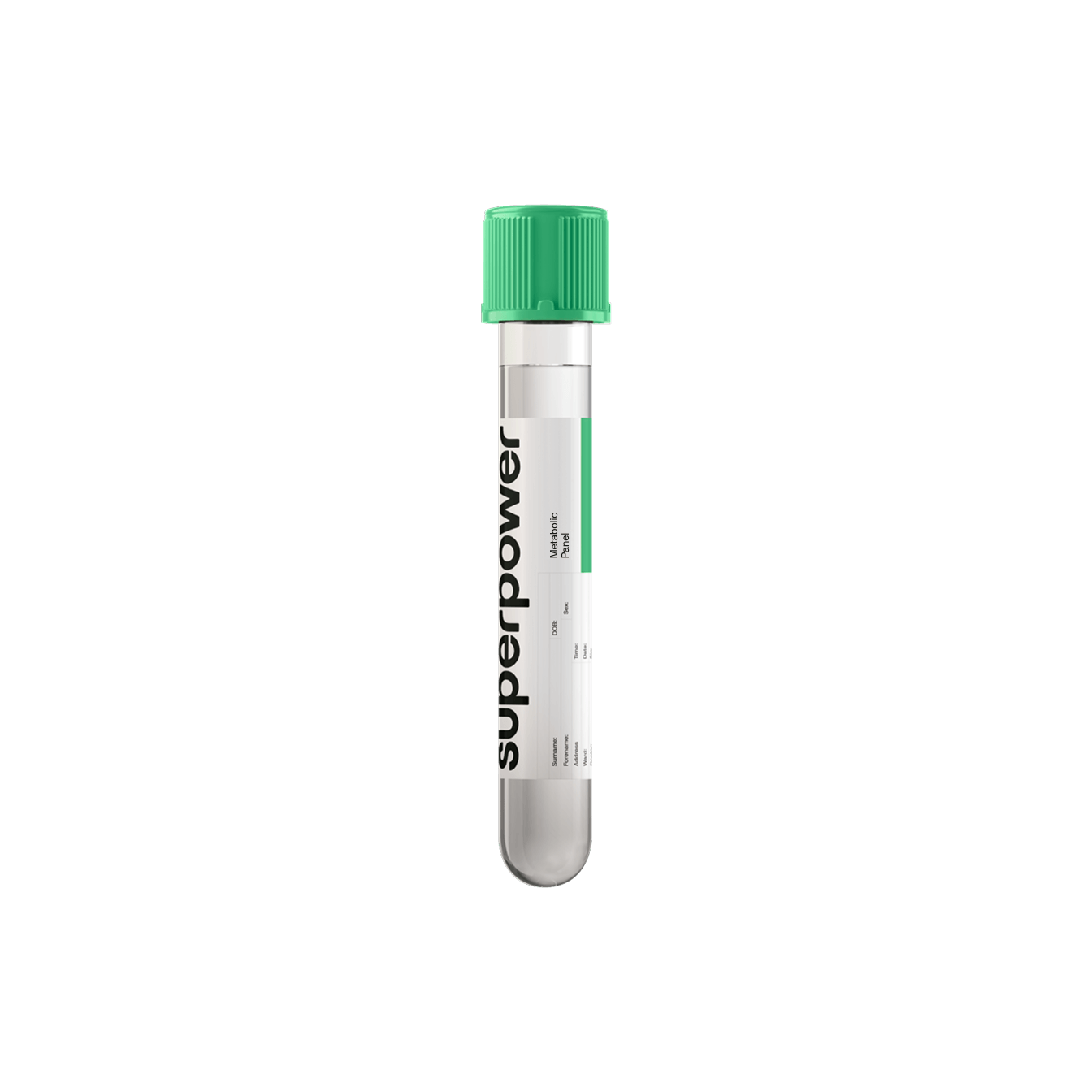
Test details
- Sample type:
- Single blood draw (blood only)
- Location:
- In-person at local lab / At-home phlebotomist visit (+$119)
- Availability:
- Available in 24 states and Washington, D.C. (not offered in NY or NJ)
- Turnaround:
- Results typically available within 10 days
- Preparation:
- 10-hour fast, water onlyNo alcohol or heavy, fatty meals the day before
About the Alzheimer's Amyloid Plaque Screening
The Alzheimer's Amyloid Plaque Screening measures amyloid protein fragments in a blood sample. It reports how the two forms stand relative to each other. That relationship, the amyloid beta 42/40 ratio, tracks amyloid status seen on brain imaging. The panel does not image your brain, and it does not count plaques.
One blood draw covers it, in-lab or at-home, and all three results come from that single sample. What comes back is information about the likelihood of amyloid pathology. You can go through it with your clinician and your Superpower care team alongside the rest of your health picture.
What this panel measures
The amyloid beta 42/40 ratio is the relationship between two amyloid protein fragments measured in the same blood sample. Amyloid beta 40 and amyloid beta 42 are each reported as their own value. The ratio is calculated from the two.
| Marker | What it reflects |
|---|---|
| ABeta 42 | The amyloid fragment that clumps together to form plaques between neurons |
| ABeta 40 | The second amyloid fragment, measured as the comparison value; on its own it may not distinguish amyloid-positive from amyloid-negative people |
| ABeta 42/40 Ratio | The relationship between the two fragments, associated with brain amyloid status |
All three values appear together on your finalized report.
Why this panel reports a ratio, not a single amyloid number
A single amyloid value is hard to read on its own, because absolute levels differ from person to person. What carries the information is the balance between them. Measured alone, amyloid beta 40 may not separate amyloid-positive from amyloid-negative participants, while the ratio does.
The ratio is not an image, not a count of plaques, and not a standalone answer. A blood amyloid result is taken into consideration along with other tests rather than read alone.
How blood, PET, and spinal fluid testing differ
Blood, spinal fluid, and brain scans are all used in an Alzheimer's workup. They differ in what is collected and in what comes back.
| Approach | How the sample or image is taken | What it produces |
|---|---|---|
| Blood | A standard blood draw | Measured amyloid fragment levels and a calculated ratio |
| Amyloid PET | A brain scan at an imaging facility | Images showing amyloid in the brain |
| Cerebrospinal fluid | Fluid collected through a spinal tap | Measured amyloid protein levels in spinal fluid |
Blood-based amyloid testing sits inside a clinician's workup. Guidance ties its use to evaluating people with objective cognitive impairment in specialized care.
What happens after a flagged amyloid result
A shifted ratio starts a clinical conversation rather than ending one.
- A clinician reviews the result against your history and the rest of your health picture.
- You discuss what the finding means for you and whether further assessment fits your situation.
- A referral to a memory specialist for further assessment can follow.
- The lab report travels with that referral, so the specialist reads your result in full.
Who benefits from testing
These are the situations members are usually in when they ask about amyloid testing.
- Alzheimer's runs in your family, and you want to know whether amyloid changes are already underway.
- You have questions about your cognitive health and would rather have objective information than a hunch.
- You want a blood-based option alongside imaging-based assessment.
- You are already discussing memory changes with a clinician — Whether the test fits you is a clinical decision. Guidance scopes blood amyloid testing to the workup of people with cognitive impairment.
What your results reveal
Your report shows the measured amyloid beta 40 and amyloid beta 42 values and the calculated 42/40 ratio. A ratio inside the expected range is reassuring information. A shifted ratio points to a higher likelihood of amyloid accumulation, for a clinician to assess. The plasma ratio predicts brain amyloid-PET status. It is not a diagnosis of Alzheimer's disease. Nor does it establish on its own that symptoms will develop.
Performance varies between blood-based biomarker tests. Take the tests reviewed in specialized care at a single cut-point. Pooled sensitivity ranged from 49.3% to 91.4%. Specificity ranged from 61.5% to 96.7%. That spread is one reason a result is read alongside the rest of the clinical picture.
Reference ranges vary by lab and individual. Your Superpower care team and your provider can help you interpret your specific results in context.
How to prepare
Follow the blood-draw instructions that come with your appointment.
- Fast for 10 hours before the draw, water only.
- Skip caffeine before the draw.
- Stay hydrated.
- No alcohol or heavy, fatty meals the day before.
- Pause supplements for 72 hours.
- No strenuous exercise for 48 hours.
- Keep taking prescribed medication unless the prescribing clinician advises otherwise, and ask your care team about any clinical preparation exception.
How it works
- Order online — Choose the draw that suits you, in-lab or at home with a phlebotomist.
- Complete your blood draw — Follow the preparation instructions, and the three amyloid results all come from that one sample.
- Review your results — Your finalized report appears in the Superpower dashboard approximately 10 days after the draw, released as a complete panel rather than in parts. Your care team can talk you through what it describes.
Frequently asked questions
Biomarkers tested
The AB42/40 ratio is a calculated value derived from your plasma amyloid beta 42 and amyloid beta 40 levels.
Learn moreMethod: Liquid chromatography-tandem mass spectrometry (LC-MS/MS) on plasma. Laboratory-developed test validated under CLIA; not cleared or approved by the FDA. Results are interpreted by clinicians in context and are not a stand-alone diagnosis.


























 Heart & Vascular Health
 Metabolic Health
 Energy
 Liver Health
 Kidney
 Sex Hormones
 Thyroid Health
 Nutrients
 Immune System
 Inflammation